


Zhang Ring Test™ Validation Across Retinal Pathologies
Clinical Validation of the Zhang Ring Test™
Correlation of Zhang Ring Test™ Findings With Clinical Diagnoses and Visual Field Defects — Dr. Fuxiang Zhang, MD
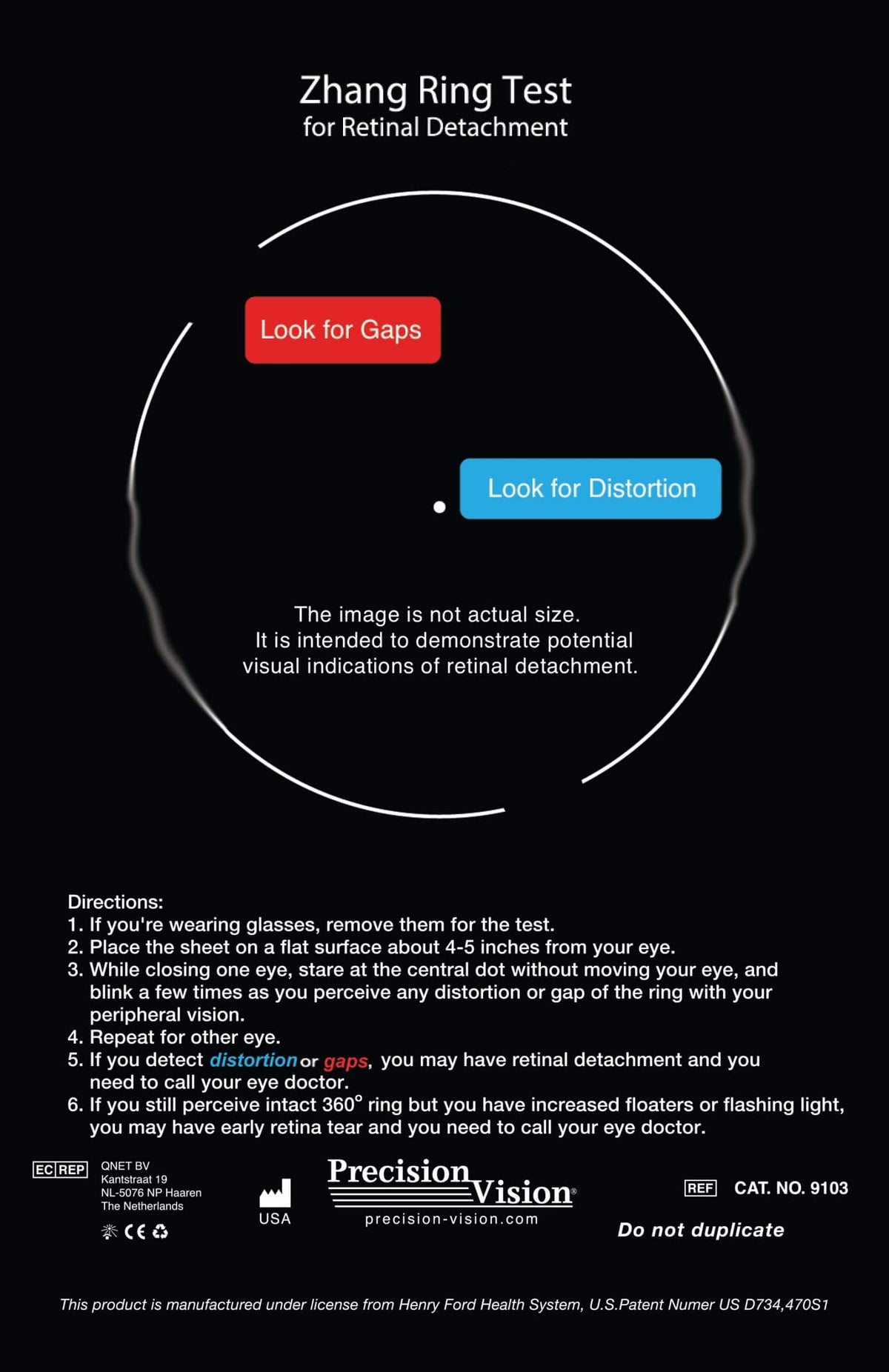
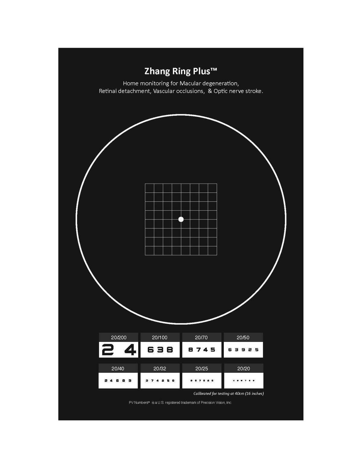
The clinical study described in this article was conducted using the original Zhang Ring Test™, the first version of the device developed to help patients identify and communicate perceived visual-field defects. The product has since been further developed and is now available as the Zhang Ring PLUS™. Accordingly, the findings and images presented in this study specifically refer to the original Zhang Ring Test™ version.
Table 3 presents a detailed analysis of 14 clinical cases involving 13 patients and 14 eyes in which patient-reported defects using the Zhang Ring Test™ were compared with conventional diagnostic modalities, including HVF, fundus photography, and physician notes. The primary objective was to assess whether findings from the patient Zhang Ring Test™ matched the clinical diagnosis and associated field defects documented through standard ophthalmic examinations.
All 14 cases demonstrated strong agreement between the Zhang Ring Test™ results and conventional diagnostic methods, supporting the potential value of patient-drawn defects and subjective visual reports when interpreted in clinical context. Several cases involved retinal detachment (RD), such as Case 1 and Case 2, where retinal detachments in the inferior and superior retina, respectively, were matched by corresponding superior and inferior field losses. In Case 3, a history of supratemporal RD treated with laser was matched with an inferior defect on the patient’s drawing and HVF, consistent with anatomical expectations due to retinal inversion in field perception.
Optic nerve pathology was also accurately reflected in several cases. For example, Case 4 involved advanced open-angle glaucoma with superior rim loss, and Case 5 involved anterior ischemic optic neuropathy (AION) with inferior nerve edema, both of which showed strong concordance among fundus findings, patient drawings, and Humphrey field test results.
Vascular pathologies such as branch retinal vein occlusion (BRVO), and branch or central retinal artery occlusion (BRAO/CRAO) were also represented. In Case 6, the patient had a BRVO with temporal hemorrhage, and the field defect presented nasally, consistent with the visual field map. Case 8 presented with acute BRAO and matching supranasal field losses. Case 9, with CRAO, revealed only a central island of vision, which was accurately described by the patient.
Two additional cases (13 and 14) involved the same patient with post-stroke visual field defects (supratemporal and supranasal quadrantanopia) that were not captured with fundus photography but aligned well with HVF and the Zhang Ring Test™ drawings, highlighting the potential application of the Zhang Ring Test™ in neuro-visual assessments.
In summary, Table 3 illustrates that there was a consistent correlation between the Zhang Ring Test™ results and standard diagnostic findings across the cases presented. These examples support the potential utility of the original Zhang Ring Test™ as a low-cost, patient-driven tool for identifying and communicating perceived visual-field changes, particularly in situations where conventional imaging or testing may be delayed or unavailable. The current product is the Zhang Ring PLUS™, which represents the subsequent version of the Zhang Ring Test™.
Table 3. Samples of Matched Cases Among the Zhang Ring Test™ and Conventional Tests.
Please pay special attention to the fourth column, “Patient Drawing.” This column provides examples of what patients with a variety of retinal and optic nerve conditions actually experienced with their affected eye. If your eye were to develop a problem such as a retinal detachment, retinal vascular occlusion, or optic nerve disorder, you might notice similar changes when viewing the Zhang Ring Test. These examples are intended to help illustrate the types of visual changes that may occur and alert you to seek appropriate eye care.
Case |
Diagnosis |
Fundus Photo |
Patient’s |
Defects on HVF or Doctor’s Note or Other Conventional Recordings |
Matching Relationship |
1 |
Right Inferior Retinal Detachment |
|
|
|
RE inferior RD with superior field missing. |
2 |
Left Superior Retinal Detachment |
 |
 |
Confrontation
|
LE superior RD with inferior field missing. Confrontation field also showed inferior missing in EMR note. |
3 |
History of Supratemporal RD and S/p laser RE |
 |
 |
 |
History of supratemporal RD and S/p laser RE with patient drawing showing infratemporal defect. HVF shows inferior defect in RE. |
4 |
Right Advanced Open Angle Glaucoma |
 |
 |
 |
Right Advanced Open Angle Glaucoma. Superior optic nerve rim is gone. All three matched well in location. |
5 |
Right AION, Sudden Loss of VA |
 |
 |
 |
RE inferior optic nerve edema with superior field loss. All three matched well in location. |
6 |
Left BRVO with lots of hemorrhage |
 |
 |
Confrontation
|
LE acute BRVO with lots of hemorrhage temporally, while field defects were mainly showed nasally. All three matched well in location. |
7 |
Right Acute BRAO with HHP |
 |
 |
 |
RE supratemporal BRAO with HHP while field defects showed inferiorly. All three matched well in location. |
8 |
Acute Left BRAO |
 |
 |
 |
LE acute BRAO with field loss mainly in supranasal area. All three matched well in location. |
9 |
Left CRAO |
 |
 |
 |
LE CRAO with small island of central tunnel vision only. All three matched well in location. |
10 |
Right ARMD |
 |
 |
 |
RE central ARMD. Patient drawing showed central fixation dot blurry while there was no problem for the peripheral ring image. |
11 |
Left Optic Pit |
 |
 |
 |
LE inferior optic nerve pit with superior field loss. All three matched well in location. |
12 |
Left Retinal Detachment with Gas Bubble at time of test |
 |
 |
 |
Left Retinal Detachment. The Gas Bubble was at superior, resulting in inferior field distortion at time of test. |
13 |
s/p stroke, |
No fundus photo taken |
 |
 |
HVF missing location matched well with patient Zhang Ring Test™ drawing. |
14 |
Same patient as #13, Left eye supranasal quadrantanopia |
No fundus photo taken |
 |
 |
HVF missing location matched well with patient Zhang Ring Test™ drawing. |


Abbreviations: RE: right eye; LE: left eye; EMR: electronic medical record; HVF: Humphrey visual field; AION: anterior ischemic optic neuropathy; BRVO: branch retinal vein occlusion; BRAO: branch retinal artery occlusion; HHP: Hollenhorst plaque; CRAO: central retinal artery occlusion; ARMD: age-related macular degeneration; PVD: posterior vitreous detachment.